Microscopy & microtechniques
Researchers in Göttingen have developed a dedicated microfluidics system that improves reproducibility and accessibility in multiplexed super-resolution microscopy, allowing scientists to visualise many cellular structures at once
A team of researchers in Germany has developed a specialised microfluidics platform that simplifies multiplexed super-resolution microscopy, an advanced imaging approach that allows scientists to visualise many molecular structures within a single cell at once. The system aims to address longstanding technical barriers that have limited the widespread use of this powerful technique.
To understand how a cell functions, scientists must examine how its internal components organise themselves and interact in a coordinated manner. Specialised cellular structures form intricate networks that operate together to control processes such as signalling, metabolism and structural stability. For researchers to map these interactions accurately, they must observe many targets simultaneously and determine how these structures relate spatially inside the cell.
Conventional light microscopy cannot resolve sufficient detail because its resolution remains limited by the diffraction of light. Super-resolution microscopy has transformed cell biology by allowing scientists to visualise structures far smaller than the traditional optical limit. In this case, the development of multiplexed super-resolution microscopy extends this capability further. It allows researchers to label and image multiple molecular targets sequentially within the same cell, thereby creating detailed maps of cellular architecture and protein interactions.
Despite its scientific value, multiplexed super-resolution microscopy often remains technically challenging. Many current workflows rely on complex laboratory procedures that require repeated manual handling steps. Small variations in how reagents enter or leave the imaging chamber can influence the outcome of the results. Fragile biological samples can also suffer mechanical stress during these procedures which reduces reproducibility and limits the types of cells researchers can examine.
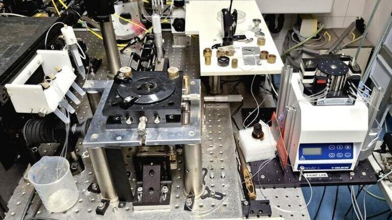
An international research team led by the University of Göttingen and the University Medical Center Göttingen, Germany, has set out to address these challenges. The researchers worked within the Göttingen Cluster of Excellence ‘Multiscale Bioimaging: From Molecular Machines to Networks of Excitable Cells’ (MBExC) to design a microfluidics system that standardises fluid handling during complex imaging experiments.
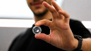
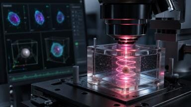
The system replaces manual pipetting with precise microfluidic control that injects and removes solutions within the sample chamber in a controlled and reproducible manner. This approach allowed researchers to perform the repeated labelling and washing steps required for multiplexed imaging without introducing variability between cycles.
“The system we have developed means we can maintain high image quality throughout long imaging cycles,” said Dr Samrat Basak, who was the joint first author of the study, and a postdoctoral researcher, who has subsequently moved to Ludwig Maximilian University of Munich.
“By keeping conditions consistent across the different labelling and washing steps, the microfluidics platform allows information from different targets to be directly mapped, making it possible to image proteins, specialised structures and complex interactions within the cell,” Basak said.
To demonstrate the capabilities of the system, the researchers carried out experiments in human cancer cells. These tests revealed the organisation of protein filaments that form part of the structural framework inside the cell. Because multiplexed imaging allows scientists to examine multiple molecular features in a coordinated manner, the approach can provide insights that single-target experiments cannot deliver.
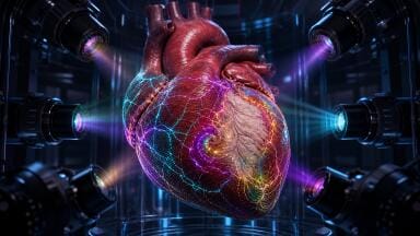
The team also applied the technology to isolated ventricular cardiomyocytes from mouse hearts. These highly specialised muscle cells generate the force that allows the heart to pump blood through the body.
“[However], the fragile, specialised muscle cells of the heart are particularly challenging to image,” said joint first author Kim-Chi Vu of the University Medical Center Göttingen and the MBExC research cluster.
“The microfluidics system was essential to complete the imaging without deforming the cells or detaching them from the surface,” she added.
According to the researchers, careful fluid control proved critical to protect delicate biological structures. Traditional manual solution exchange can create local disturbances within the imaging chamber. Even minor mechanical stress can distort cell morphology or disrupt cellular components during the long experimental sequences required for multiplexed microscopy. The microfluidic platform minimises these disturbances and ensures that reagents enter and leave the chamber in a stable and predictable way.
The system also offers flexibility for different research environments. Scientists can operate the device either manually or through automated control, which allows laboratories to adapt the platform to different experimental needs. Importantly, the design remains compatible with a wide range of existing microscopy systems which could help laboratories adopt the approach without major changes to their equipment.
“The core idea was to develop a system which is cost-efficient, adaptable, and can be redesigned according to specific imaging needs of complex biological systems,” said Dr Roman Tsukanov, a senior postdoctoral researcher at the University of Göttingen.
“By automating fluid exchange, we removed a major source of variability and made complex imaging protocols much more user-friendly,” he said.
Researchers believe that improved accessibility could allow multiplexed super-resolution microscopy to play a larger role across cell biology, biomedical research, and translational medicine. Detailed maps of molecular organisation inside cells could help scientists investigate how diseases alter cellular architecture and identify potential therapeutic targets.
“This approach will help standardise multiplexed super-resolution imaging and make it broadly accessible, benefiting both research and medical applications,” said Professor Jörg Enderlein of the University of Göttingen’s Faculty of Physics.
The authors suggested that standardisation and automation represent essential steps for the broader adoption of advanced microscopy techniques. As laboratories continue to seek ways to map cellular processes with greater precision, tools that improve reproducibility and protect fragile samples may prove crucial to unlock the full potential of super-resolution imaging in biological research.
For further reading please visit: 10.1021/acsnano.5c18697
ILM 51.6 Sept 2026





 Product directory