Microscopy & microtechniques
Harvard University researchers have developed a cathodoluminescence-based multicolour electron microscopy technique that enables simultaneous protein localisation and ultrastructural imaging in cells at nanometre scale
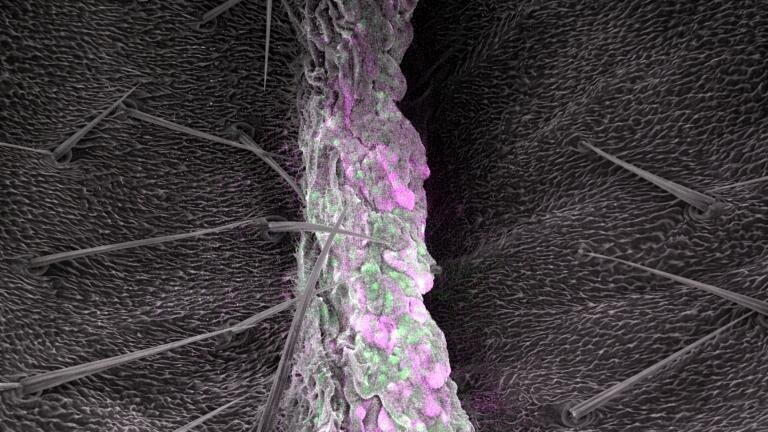
Scientists have developed a novel imaging technique that unites the molecular specificity of fluorescence microscopy with the ultrastructural power of electron microscopy and has enabled researchers to visualise both the fine architecture of cells and the precise location of proteins in vivid colour at nanometre resolution. The advance – known as multicolour electron microscopy – has addressed a longstanding challenge in biological imaging, where investigators have had to choose between structural detail and molecular identification but could not readily achieve both in a single experiment.
The work has opened opportunities to study cellular signalling pathways, protein clustering and subcellular organisation while retain structural context.
“I’ve always been fascinated by developing microscopy techniques that can image things we haven’t seen before,” said Dr. Debsankar Saha Roy, a fellow in the laboratory of Maxim Prigozhin at Harvard University.
“We’re building a multicolour electron microscope – a technique that combines [both] the benefits of electron microscopy and fluorescence microscopy,” he said.
Conventional fluorescence microscopy relies on fluorescent tags that bind to proteins of interest. When visible light illuminates the specimen, these tags emit light and thereby mark the location of specific molecules. This approach has proved powerful for molecular localisation but its spatial resolution is fundamentally limited by the diffraction of light. At that scale, individual proteins cannot resolve and, on the other hand, fluorescence images provide little information about the broader cellular landscape beyond the labelled targets.
“[Due to the diffraction of light] resolution is limited to about 250 to 300 nanometres, so you can’t see individual proteins clearly,” Roy explained.
“But the bigger issue is that you don’t see the structure of the cell. You see whatever is labelled but you don’t see everything else around it,” he added.
Electron microscopy, by contrast, can reveal cellular ultrastructure down to a few nanometres, orders of magnitude finer than light microscopy. It has enabled scientists to map membranes, organelles and macromolecular assemblies in extraordinary detail. Yet traditional electron microscopy has struggled to identify specific proteins in colour within those complex landscapes. Attempts to combine the two modalities have typically required separate imaging sessions followed by digital overlay. Precise alignment, particularly in large or heterogeneous samples such as brain tissue, has often proved unreliable.
The Harvard team adopted a different strategy. Instead of separate optical and electron imaging, they have used a single electron beam to generate both structural and molecular information at once.
“We’re not sending in light – we’re sending an electron beam,” Roy said.
“We have probes that you can attach to a protein that emit visible light when excited by electrons. This process is called cathodoluminescence.
“So, from the same electron beam, you get two sets of information: the coloured signal from the probes and also the detailed structural image from the electrons,” he said.
Cathodoluminescence refers to the emission of photons from a material after excitation by an electron beam. In this system, electron excitation produces a colour signal from labelled proteins while the scattered electrons simultaneously form a high-resolution structural image. Because both signals originate from the same excitation source, spatial correspondence remains intrinsically aligned at nanometre scale.
An important advantage of the approach lies in its compatibility with established labelling tools. The team had previously developed lanthanide nanoparticles as probes for multicolour electron microscopy and had worked to attach them to proteins. More recently, they observed an unexpected phenomenon when they introduced common fluorescent dyes into the electron microscope.
“The most surprising thing we observed was that standard dyes used in fluorescence microscopy also emit visible light when you excite them with electrons,” Roy said.
“That had never been seen before. And these dyes – and their protein labelling methods – are already developed and available. So, you don’t have to create anything new,” he said.
This finding suggests that laboratories can adapt widely available fluorescent dyes for cathodoluminescence without the need to engineer entirely novel reagents. The possibility to repurpose established probes has the potential to facilitate uptake and reduce technical barriers.
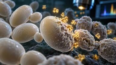
The researchers have already demonstrated the method in mammalian cells and in biological tissues, including fungus-infected flies, which provided a complex and physiologically relevant test system. The technique has therefore moved beyond proof-of-principle and has begun to show practical utility in diverse biological contexts.
At present, the platform only generates two-dimensional images and so the next objective is to extend the method into three dimensions (3D), particularly through integration with cryo-electron microscopy. In cryo-electron microscopy, rapid freezing preserves cells in a near-native state and allows researchers to collect multiple projections for 3D reconstruction.
“We want to extend this multicolour electron microscopy approach to 3D. To get there, we aim to implement this technique in ultrathin sections of cell embedded matrices and or in cryo-electron microscopy – that’s the next step,” Roy concluded.
If successful, a 3D multicolour electron microscopy platform could allow scientists to map the molecular identity of proteins within intact cellular volumes at nanometre precision. Such capability would provide a powerful tool to dissect the spatial logic of signalling complexes, organelle organisation and macromolecular assemblies within their native structural environment.
By uniting molecular specificity with ultrastructural context in a single instrument, multicolour electron microscopy has the potential to reshape how researchers interrogate cells.
ILM 51.6 Sept 2026




 Product directory