News
Published over 6 years ago. See the latest and most current information on News.
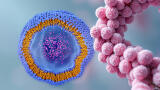
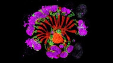
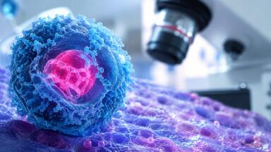
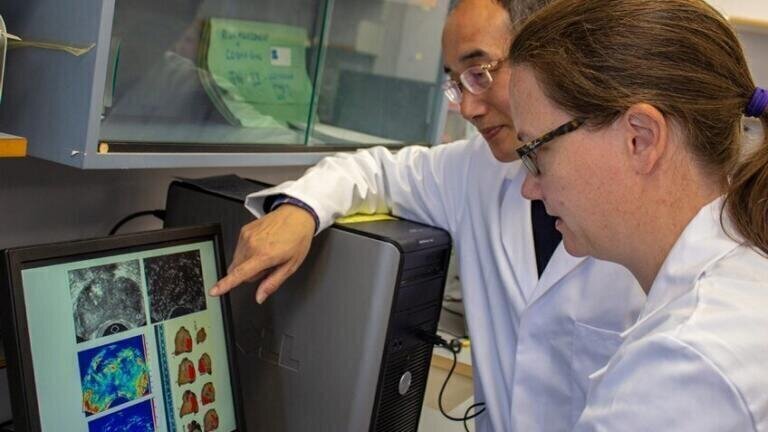
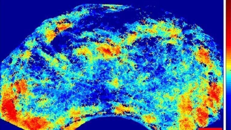
A new technique using super-resolution ultrasound methods for scanning whole organs has been discovered by a research team at the Heriot-Watt University, Edinburgh. The technique, found to have improved resolution by 5-10 times compared to standard ultrasound images, demonstrated for the first time the detection of prostate cancer by mapping the blood vessels that surround it and showing a different pattern to that of normal tissue. The discovery has the potential for earlier cancer diagnoses, allowing medical staff to more effectively target treatments to any malignant tissue.
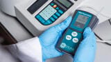
The enhanced images utilise existing clinical two-dimensional (2D) ultrasound equipment and standard Contrast Enhanced Ultrasound (CEUS) modes. This means hospitals won’t be required to invest in new equipment and no new hardware technology needs to be developed. The aim is to start human trials using the new technique later in 2019. Prostate patients will be the first to benefit from the enhanced imaging.
The breakthrough uses adaptive optics, first applied in astronomy and then (under STFC funding) was successfully adapted for use in optical microscopy, leading to the Heriot-Watt team developing micrometric resolution ultrasound imaging.
Lead researcher Dr Vassilis Sboros explained: “Ultrasound imaging is an indispensable tool in medical diagnosis, primarily due to its cost-effectiveness and unique real-time capability. However, the limitations of current ultrasound images mean more expensive techniques like MRI are often employed for diagnosis and treatment.
“MRI doesn’t provide clinicians with more detail but it has generally provided better results than other methods. However, in the prostate for example, biopsy has to be performed as a separate procedure which is more expensive for the hospital and can be both disruptive and distressing for the patient.
“Our new technique has the advantage that it can be done as an adjunct to the ultrasound examination which allows the biopsy to be integrated into it. Due to the super-resolution capability of the image created, we anticipate that the ability of the medical staff to pinpoint, diagnose and treat a range of cancers will be greatly enhanced.”
Published in the Journal of Investigative Radiology
ILM Guide 2026/27

.jpg)