Clinical, medical and diagnostics
A non-destructive imaging technique has revealed how metabolically active individual immune cells are within a single blood sample, opening new possibilities for diagnostics and cell therapy manufacturing
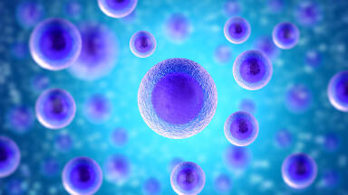
Each routine blood sample contains a wide range of immune cells that can be used to reveal important clues about a patient’s health or their disease. These cells – known as peripheral blood mononuclear cells (PBMCs) – are widely used to study infections, autoimmune disorders, cancer and the immune system’s response to treatment. Scientists have previously identified these cells by use of fluorescent labels that attach to specific surface markers. While effective, those methods provide limited information about how the cells function and can alter them during preparation.
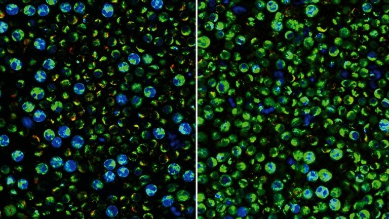
A recent study has now shown how advanced optical imaging can reveal a previously inaccessible layer of information which is the metabolic activity of individual immune cells within a complex blood sample.
“PBMCs can be isolated clinically really easily, and they’re already used in the clinical workflow. So, the question is, what can we get from them that we aren’t already getting?” said Dr. Melissa Skala, who leads the Skala Lab and is senior author of the study. She is also the ‘Carol Skornicka Chair’ at the Morgridge Institute for Research and professor of biomedical engineering and medical physics, which is part of the University of Wisconsin–Madison, Madison, USA.
PBMCs are routinely studied in conditions ranging from blood cancers and sepsis to lupus and cognitive decline. They also serve as the starting material for cell therapies such as chimeric antigen receptor T-cell (CAR T) treatments which can engineer a patient’s own immune cells to attack cancer. Understanding how many immune cells are present, and how active and metabolically fit the components are, could provide new insights into disease progression and treatment response.
Up until now immune-cell metabolism could only be assessed through the isolation of specific cell populations or through the addition of fluorescent labels or chemical probes. These approaches can alter cells, consume samples or fail to capture how each different cell type interacts with others. This study has demonstrated a non-destructive method to measure metabolism in individual immune cells while they remain part of a heterogeneous PBMC sample.
The researchers achieved this using optical metabolic imaging (OMI) – a technique developed and refined by the Skala Lab. OMI relies on the natural fluorescence of molecules involved in cellular energy production. Rather than introduce any external dyes, their method uses two-photon microscopy to excite naturally occurring metabolic cofactors inside cells and to measure how long they emit light. These fluorescence lifetimes provide information about cellular metabolism and activation state. Because the approach uses endogenous signals, the cells remain intact and available for further analysis and could therefore ultimately have a therapeutic use.
The team applied OMI to PBMC samples isolated from the blood of three healthy donors, then analysed thousands of individual cells in both resting and activated states. Machine-learning algorithms were used to determine whether metabolic measurements alone could identify different immune-cell populations and detect immune activation.
The results showed that metabolism provides a powerful indicator of immune-cell identity and function. The researchers were able to distinguish activated from resting PBMCs with nearly 94 per cent accuracy just two hours after stimulation. They also identified monocytes – key cells of the innate immune system – with a 96 per cent level of accuracy at rest and 88 per cent accuracy in activated samples. Natural killer (NK) cells were identified with approximately 74 per cent accuracy in both states.
The findings highlight the importance of immune-cell metabolism. Although immune-cell counts are already used as diagnostic markers, metabolism can offer additional information about whether cells are active, quiescent or responding to a threat. The study found that monocytes and NK cells have particularly distinct metabolic signatures, which is likely to reflect their role as rapid responders in the immune system. T cells and B cells, which drive more specialised adaptive immune responses, displayed more similar metabolic profiles under the experimental conditions.
Importantly, the single-cell approach revealed substantial heterogeneity within the immune-cell population. Some cells, such as monocytes, showed high levels of metabolic activity, while others remained quiet. Such differences would be obscured by bulk measurements that average signals across an entire sample. By measuring metabolism at the level of individual cells, OMI offers a more detailed picture of how the immune system functions.
The non-destructive nature of the technique could prove especially valuable for cell therapy applications. Current methods to assess cellular metabolism often require reagents or processing steps that can alter cell behaviour. OMI, by contrast, leaves cells viable after analysis, which creates opportunities to evaluate cell quality before the manufacture of therapeutic products. As PBMCs are commonly used as starting material for CAR T-cell therapies and other immune-cell treatments, pre-treatment assessment of cellular fitness is an attractive possibility.
The team emphasised that the technology remains primarily a research tool and does not yet match the accuracy of established labelling methods for the identification of every immune-cell subtype. However, its key ability to measure single-cell metabolism without sample destruction could make it a valuable complementary analysis tool alongside existing approaches.
For further reading please visit: 10.1117/1.BIOS.3.3.035003
ILM 51.6 Sept 2026



 Product directory


.jpg)