Laboratory products
Published over 8 years ago. See the latest and most current information on Laboratory products.
As the highest honor in science, Nobel Prizes are awarded to individuals who pioneer exceptional academic, cultural or scientific advances. This year, a trio of researchers have won recognition in the chemistry category for their roles in developing cryo-electron microscopy.
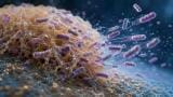
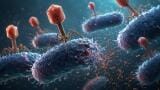
Their achievement? Astoundingly detailed 3-D snapshots of life’s atomic details. Known as cryo-electron microscopy, the ultra-advanced imaging technique essentially freezes tiny biological objects like proteins and viruses in place, which allows scientists to explore their structures at the scale of atoms.
Together, Jacques Dubochet of Switzerland's University of Lausanne, Joachim Frank of America's Columbia University and Richard Henderson of England's MRC Laboratory of Molecular Biology won the 2017 Nobel Prize in Chemistry for their contributions to the development of cryo-electron microscopy.
“Now that we can see the intricate details of every drop of our body fluids, we can understand how they are built and how they act and how they work together,” applauds Sara Snogerup Linse, chair of the 2017 chemistry Nobel committee. “We are facing a revolution in biochemistry…. Soon, there are no more secrets.”
In the past structural biologists have relied on nuclear magnetic resonance (NMR) spectroscopy and X-ray crystallography to map the microscopic landscape of molecules. While they are effective, both techniques have limitations. For example, the latter bounces X-rays off a crystallised version of a molecule to create a 3-D picture. However, as a lot of proteins cannot be crystallised their structures remain inaccessible.
Cue cryo-electron microscopy, also known as cryo-EM. It was first pioneered in the 1970s, when Henderson attempted to use regular electron microscopy to study proteins embedded in cell membranes. Though he was unable to crystallise the protein as removing it from the membrane could compromise its structure.
In 1984, Jacques Dubochet built on the concept and used a thin layer of flash frozen “vitrified water” to protect his samples from dehydration in electron microscope vacuums. This technique is now a crucial component of modern cryo-electron microscopy.
A series of improvements went on to advance cryo-electron microscopy to the atomic scale. The use of a computer algorithm was introduced by Frank, allowing scientists to scan images, detect proteins oriented in the same direction and group them together. In 1990, Henderson produced the first cryo-EM image of a protein at the atomic scale.
Over the past few years cryo-EM has begun to flex its muscles, and is now offering a glimpse at the structure of larger objects like viruses. In fact, in 2016 the technique was used to map the structure of the Zika virus.
Cryo-EM has opened exciting new doors for structural biology, with experts hailing it as the "resolution revolution." Next up? Mapping entire cells.
Spectroscopy is an incredibly complex field, with laboratories relying heavily on sophisticated equipment. For a closer look at the industry 'Compliance, CRMs and Accreditation in Spectroscopy – A Supplier’s Perspective' offers a glimpse at how regulators and commercial suppliers have met the demand for reliable reference materials.
ILM 51.5 July 2026


.jpg)
.jpg)


-(1).jpg)
.jpg)
.jpg)
.jpg)
.jpg)
.jpg)