Laboratory microscope
Technique combines structured illumination with mid-infrared photothermal detection to give chemical imaging at high-speed
The current generation of super-resolution microscopes can make observations at nanoscale with extraordinary precision. Fluorescent tags are required that show structural detail of study samples but give low levels of chemical information. Conversely the novel vibrational imaging technique can identify molecules based on their unique chemical bonds without altering the sample.
These methods detect physical changes in samples when they absorb mid-infrared (MIR) light, such as shifts in refractive index caused by heat absorption or temperature-induced acoustic signals. Existing methods however struggle at weak signal levels, making it difficult to achieve both high resolution and strong chemical contrast such that molecules can be distinguished.
The newly developed technique of structured illumination midinfrared photothermal microscopy (SIMIP) has been shown to address this limitation with double the resolution levels of conventional microscopy.
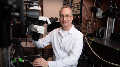
Developed at Zhejiang University, China, by a research team led by Professor Delong Zhang, their novel technique represents a significant advance in vibrational imaging, opening up new frontiers for both nanoscale chemical and biological analysis.
“SIMIP microscopy integrates the principles of structured illumination microscopy with midinfrared photothermal detection. Mid-infrared photodetection provides chemical specificity, while structured illumination microscopy enhances the spatial resolution of the sample,” said Zhang.
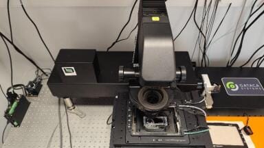
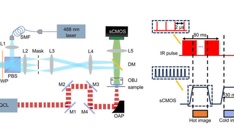
Consisting of a quantum cascade laser (QCL) that excites specific molecular bonds, the system causes localised heating that reduces the brightness of adjacent fluorescent molecules. Simultaneously, a structured illumination microscope (SIM) system consisting of a 488-nm continuous-wave laser and a spatial light modulator (SLM) generates striped light patterns that are projected onto the sample at different angles. These patterns create Moiré fringes, encoding previously unresolvable high-frequency details into detectable low-frequency signals that are captured by a scientific CMOS (sCMOS) camera.
Comparison of images taken with and without vibrational absorption, SIMIP therefore reconstructs high-resolution images rich in both chemical and spatial information.
The team achieved a spatial resolution of around as proof of concept, by applying Hessian SIM and sparse deconvolution algorithms achieve a higher spatial resolution, up to ∼60 nm, with an imaging speed of more than 24 frames per second, surpassing conventional MIR photothermal imaging.
To validate the accuracy of SIMIP, researchers tested it on 200-nm polymethyl methacrylate beads embedded with thermosensitive fluorescent dyes. By sweeping the QCL across the 1420–1778 cm-1 range, SIMIP successfully reconstructed the vibrational spectra, closely matching results from Fourier transform infrared (FTIR) spectroscopy.
In terms of resolution, SIMIP achieved a 1.5-fold improvement over conventional MIR photothermal imaging, with a full width at half-maximum (FWHM) of 335 nm versus 444 nm in standard methods. Moreover, it was able to distinguish between polystyrene and polymethyl methacrylate beads within sub-diffraction aggregates, which was impossible with standard fluorescence microscopy.
An added advantage of SIMIP is its ability to detect autofluorescence – the natural fluorescence emitted by certain biological molecules. This can be achieved by switching from widefield SIM to point-scanning SIM for structured excitation of autofluorescence or by using a shorter-wavelength probe beam for a widefield photothermal detection method to enhance compatibility with existing optical setups.
By integrating SIM with MIP, SIMIP achieves high-speed, super-resolution chemical imaging beyond the diffraction limit. This method opens new possibilities for observations in materials science, biomedical research, and chemical analysis. For example, the researchers envision using SIMIP to detect small-molecule metabolites and analyse their interactions with cellular structures.
The team now plans to enhance SIMIP’s temporal synchronization to further improve imaging speed and accuracy, as well as explore temperature-sensitive dyes to increase sensitivity. With minimal hardware modifications to existing SIM systems, SIMIP is poised for adoption in laboratories worldwide.
For further reading please visit: 10.1117/1.AP.7.3.036003
ILM 51.6 Sept 2026




 Product directory